
Unmasking “zombie cells” in aging tissue with an AI-powered barcode
As we age, some of the cells in our body enter a state of senescence, in which they stop dividing but do not die. Those senescent cells can contribute to age-related disorders such as cancer, tissue degeneration, and inflammatory diseases.
In an advance that could lead to better ways to diagnose and treat those diseases, MIT researchers have developed a noninvasive way to detect biomarkers of senescence. Their method is based on Raman microscopy, which can reveal the biochemical composition of cells without harming them.
By combining Raman microscopy with gene expression data at single-cell resolution from the same cells, the researchers were able to identify unique “barcodes” that can be used to quickly identify senescent cells. This study was done in mouse cells, but the researchers are now working on adapting it for use with human tissue.
“You can imagine that one day we may develop an endoscope that can look inside your body and identify cellular senescence,” says Jeon Woong Kang, an MIT research scientist and one of the senior authors of the study.
The research is part of a National Institutes of Health initiative called the Cellular Senescence Network, which is pursuing a deeper understanding of senescence in hopes of developing therapies that could combat some of the tissue-damaging effects of senescent cells.
Peter So, director of the MIT Laser Biomedical Research Center (LBCR) and an MIT professor of biological engineering and mechanical engineering, and Jian Shu, an assistant professor at Massachusetts General Hospital (MGH) and Harvard Medical School, and an associate member of the Broad Institute and Ragon Institute, are also senior authors of the paper, which appears today in Nature Aging. Lead authors of the paper are Ke Zhang, an instructor at MGH and Harvard Medical School; Xingjian Chen, a postdoc at MGH and Harvard Medical School; Francesco Monticolo, a postdoc at MGH and Harvard Medical School; and Salvatore Sorrentino, a postdoc at MIT.
Characterizing senescence
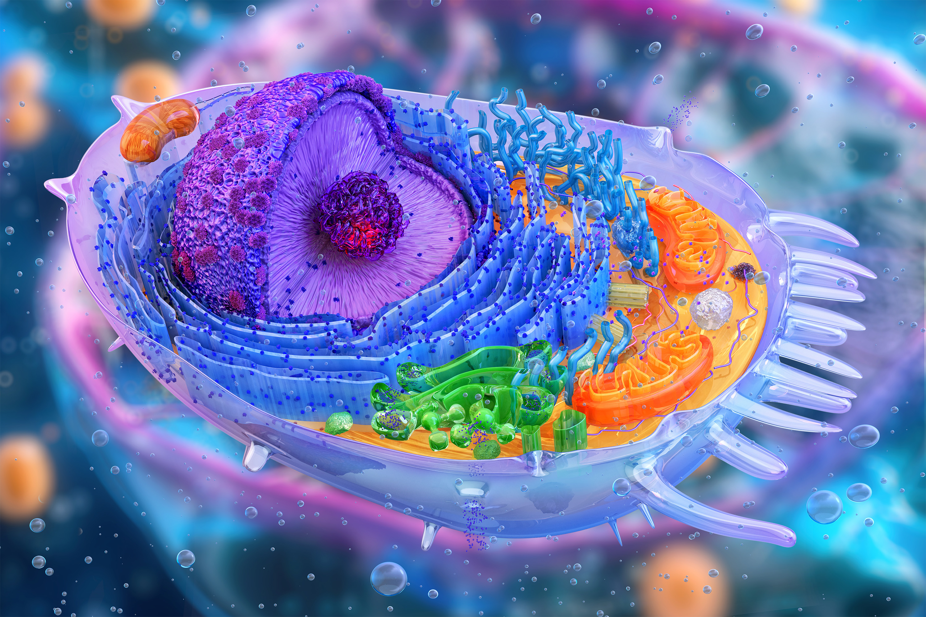
Cell senescence is often triggered by DNA damage, which leads to an irreversible arrest of the cell cycle. These cells don’t die, but they undergo significant changes to their shape, metabolic processes, and gene expression profiles.
The immune system is responsible for clearing out these “zombie cells,” but as people age, this process becomes less efficient. When senescent cells accumulate, they may contribute to sagging skin, muscle weakness, and chronic conditions such as osteoarthritis and type 2 diabetes.
Cellular senescence also has beneficial effects, playing critical roles in embryonic development and tissue regeneration.
“Senescence is not just a pathological condition,” So says. “The idea behind the NIH Cellular Senescence Network is to take a very comprehensive approach to understand senescence and identify senescent cells, because it plays a role in so many normal physiological conditions and many pathological conditions.”
Scientists have already identified a few biomarkers for senescence, including two proteins called p16 and p21, which are involved in halting the cell cycle. However, those proteins can only be identified using a process that ends up destroying the cells.
The MIT team wanted to find a way to noninvasively identify senescent cells using Raman microscopy. Unlike RNA-sequencing, which consumes the cells as it analyzes them, Raman microscopy is a nondestructive technique that reveals the chemical composition of tissues or cells by shining near-infrared or visible light on them.
In the new study, the researchers used Raman microscopy in conjunction with spatial RNA sequencing — a technique that reveals where genes are active within a tissue — to identify new markers of senescence. By combining these two techniques, they were able to generate a much broader picture of the distinctive features of senescent cells, including gene expression levels, spatial location, and other biochemical information.
In this video, Raman microscopy is used to image skin tissue. Each frame corresponds to a different wavelength of light scattered by the tissue, and brighter colors indicate a stronger Raman signal from that part of the tissue. Analyzing these signals can reveal the biochemical composition of the tissue.
Credit: Courtesy of the researchers
“Our idea was to look at many different features to characterize senescence. That’s why we wanted to combine both single-cell gene expression and Raman microscopy, so that we can characterize the senescence from two complementary views,” Shu says.
Using both methods of analysis, the researchers examined skin and lung tissue from 2-month-old mice and 26-month-old mice.
One of the most dramatic changes seen in both lung and skin cells was an increase in lipid synthesis in older cells, along with accumulation of lipids. How this affects the physiology of the cells is not yet known, the researchers say.
The researchers also found some effects that were specific to each tissue. In senescent skin cells, they discovered that cellular pathways associated with muscle contraction and with remodeling of collagen and the extracellular matrix were significantly affected. And in aged lung tissue, they found increased activity of genes involved in immune activation and inflammation.
In future work, the researchers hope to study further what role these changes play in senescent cells.
Identifying senescent cells
Using these data, the researchers were able to identify combinations of Raman peaks that correlate with senescence. These peaks, which represent specific chemical bonds, are linked to the presence of certain lipids, proteins, or other molecules.
“Combining the most important Raman features with the most important gene signatures, we were able to create a barcode that can help us to identify senescent cells in a more unbiased way,” Sorrentino says. “Using this barcode, we can focus on a few Raman bands that emerged as the most informative in this work.” Using these bands, it could be possible to identify senescent cells by looking for just those bands of the Raman spectrum. This could help to enable diagnostics that would detect cells that have become senescent.
To help make that possible, the researchers are now working on a higher-speed version of their Raman imaging system. Currently, it takes about 30 hours to analyze a tissue sample about one square millimeter in size, but they hope to develop a system that can quickly pick out the Raman barcodes they identified from larger samples.
The research was funded by the National Institutes of Health and Massachusetts General Hospital.
What did you think of this news? Leave a comment below and/or share it on your social media. This way, we can inform more people about the hottest things in technology, science, innovation, and gaming!
This news was originally published in:
Original source
