
Astronauts Have Taken The First Human X‑Rays in Space
For more than 65 years, humans have been soaring through space.
Since the first spaceflight by cosmonaut Yuri Gagarin in April 1961, the human presence in space has become a constant, with a revolving team of international spacefarers residing aboard the International Space Station.
In the coming decades, our presence in space is likely to expand – and with it, the need for vital medical tools to keep our cosmic explorers healthy.
Now, for the first time, astronauts in orbit have taken diagnostic-quality X-rays of their own bodies – the culmination of years of work. The results are now published in the journal Radiology.
“It felt historic in a number of ways,” aerospace medicine researcher Sheyna Gifford of Mayo Clinic told ScienceAlert.
“The fact that it happened changed the future of space medicine and space missions. In an instant, what had previously been impossible was made possible.”
For more than four decades, ultrasound has been the only practical medical imaging technology available to astronauts in orbit.
This technology works by propagating high-frequency sound waves into the human body and observing how they bounce back from the tissues therein. Obtaining and interpreting ultrasound images requires considerable training, but the technology is very versatile.
By contrast, X-ray imaging requires an X-ray source, a detector on the opposite side of the body, and a human patient perfectly positioned between them. You also need everything to stay still long enough for the imaging to succeed.
Ultrasound became the standard imaging tool for space because it is portable and safe, and its transducer can be pressed directly against the body, making it much easier to use in a microgravity environment where everything tends to float away.
In recent years, X-ray imaging technology has been shrinking, and with the emergence of small, portable, battery-operated devices, scientists started to believe that in-orbit X-ray imaging might be possible.
“The change would be significant: A faster, more accurate, painless diagnosis. An X-ray is one of the most powerful diagnostic tools in modern medicine because of its speed, accuracy, and ability to be operated by a broad range of people,” Gifford said.
“X-ray in space has the power to upgrade a suspected fracture to a confirmed fracture in a fraction of a minute.”
A major breakthrough took place in 2022, when scientists managed to take X-ray images aboard a parabolic flight that briefly simulates orbital microgravity.
But, having shown that the technique can work for seconds at a time, the next hurdle was seeing if it could work in an orbital context.
The setting was SpaceX’s Fram2, an all-civilian spaceflight mission on a 3.5-day polar orbital flight aboard the spacecraft Resilience. (Fram2 was named after the ship that carried renowned polar explorers Fridtjof Nansen and Roald Amundsen).
The crew carried an X-ray system that featured an ultraportable, wireless digital X-ray generator. They received four hours of training and took X-ray images of their bodies before and during the orbital flight.
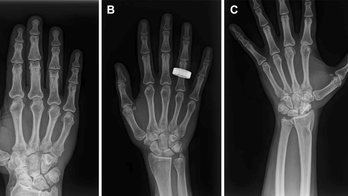
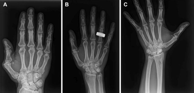
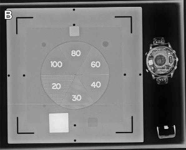
The images included a phantom object that served as a control, a smartwatch, hands, forearms, chests, abdomens, and pelvises.
All the X-ray images were independently evaluated by radiologists on Earth, who found that the scans were all of a similar high quality suitable for diagnosis.
“In 2022, we partnered with a commercial X-ray company to buy two seats on the parabolic flight provider ZeroG. When the gravity turned off, we floated up from our seats, I held up my hand, and the first digital X-ray in altered gravity was taken, just like that,” Gifford said.
“For decades, scientists were turned off by the daunting number of degrees of freedom in the belief that it would result in a blurry image. Our solution? Take the picture really, really fast.”
As expected, the greatest difficulty wasn’t obtaining the X-ray images, but positioning the patient, detector, and X-ray source and keeping them still long enough to obtain a good image.
The hands and arms were the easiest parts to image cleanly because they are easy to keep still. The images of the chest, abdomen, and pelvis proved more difficult to obtain; they were slightly lower in quality than the hand and arm images, but still above the threshold for diagnostic use.

The technology is likely not going to be limited to diagnostic use on humans, either.
As the images of the smartwatch showed, a portable X-ray machine could be used to inspect spacecraft equipment and other mission-critical hardware for hidden damage, which the researchers refer to as non-destructive testing.
“The current application of spectral X-ray on Earth started with, and remains largely used for, nondestructive testing: Its use in airport security is worldwide,” Gifford explained.
“While this team was the first to attempt spectral X-ray in space, one of the many reasons to do so is because it brings to the mission powerful tools that function far beyond the medical bay.”
There are still some limitations to be addressed. In-flight imaging time imposed a limit on the number and variety of X-ray images that could be obtained, and real-time telehealth may be unavailable for future spaceflight missions.
The researchers suggested AI-assisted analysis could eventually help astronauts assess image quality and identify potential medical problems on missions where expert radiologists on Earth are too far away to provide immediate support.

In addition, the apparatus sustained damage when the crew returned to Earth, although it remained functional. For future missions to the Moon and Mars, a more robust, rugged system may need to be developed.
Related: Exclusive Interview: Scientists Are Making New Drugs in Space
As humans prepare for those historic journeys, the ability to diagnose illnesses and injuries without relying on Earth becomes ever more important.
This research represents a critical step towards providing the fundamental medical tools that those future explorers will need as they boldly venture beyond Earth and out into the Solar System that awaits.
“On Earth, we consider this system tremendously compact and portable,” Gifford said.
“In space, this system is considered massive. For X-rays in space to become routine and for the mass and volume the system takes up to be justified, it would need to be a fraction of the volume it is now.”
She also noted that it needed to be hardened for the vacuum, and that real-time integrated support would “make this system near universal on space missions: Whether there are humans onboard or not.”
The findings have been published in Radiology.
This article was fact-checked by Carly Cassella and edited by Rebecca Dyer. While we pride ourselves on our process, we are only human. If you spot a mistake, please let us know.
What did you think of this news? Leave a comment below and/or share it on your social media. This way, we can inform more people about the hottest things in technology, science, innovation, and gaming!
This news was originally published in:
Original source
